
Ultrawide-field (UWF) imaging has changed the way we manage patients with retinal disease. However, the modality is not without its limitations. On older UWF imaging platforms, these include low resolution (which affects our ability to simultaneously image the area near the optic nerve and the periphery), lack of true color images, and the inability to capture multiple image modalities (eg, fundus autofluorescence [FAF] green, FAF blue, fluorescein angiography [FA], etc) on a single platform. In addition, lid and lash artifacts frustrate our efforts to acquire clean shots of our patients’ posterior and peripheral retinas.
The ZEISS CLARUS 700 is a single UWF platform that addresses a number of the issues clinicians encounter in their clinic. True color, FAF blue, FAF green, and FA are all available on the CLARUS 700, as are external photography and infrared imaging (Figure 1).
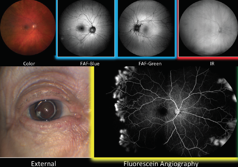
Figure 1. Multiple modalities exist in the single platform on the ZEISS CLARUS 700.
Rather than use a single white light to acquire UWF true color fundus photographs, the CLARUS 700 uses three different colored light-emitting diodes (LEDs) with broad line fundus imaging to scan the retina during color fundus photography. By using a red LED (585-840 nm wavelength), green LED (500-585 nm), and blue LED (435-500 nm), the platform provides a natural-looking fundus photograph as it appears through direct observation.
Lash and lid artifacts are removed via algorithmic adjustment, providing a cleaner image for assessment. The accuracy and resolution are those of a traditional 45° camera but provide a widefield view up to 200° with montaging.
The CLARUS 700 houses a trio of features that make the platform easier to use and boosts the data available in the clinic. PrecisionFocus technology enhances our ability to examine a particular region of interest without sacrificing resolution in another area. AutoBright software optimizes an entire series of images (rather than optimizing on an image-by-image basis) for brightness. GazePoint artificial intelligence (AI) technology facilitates montaging by identifying and using the optic nerve as a focal point, providing a superior UWF image.
PrecisionFocus
PrecisionFocus is particularly useful in non-emmetropic
eyes. In these eyes, features near the foveal center are in focus, and images in the periphery are rendered in lower resolution. With PrecisionFocus technology, details in the foveal center and the periphery are both rendered in high resolution (Figure 2).
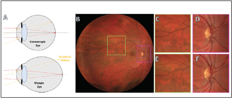
Figure 2. Peripheral defocus is a common issue encountered when imaging non-emmetropic eyes (A). Focusing on the vessels (B, yellow box) in a 90° fundus image of a myopic eye may result in less sharp images near the foveal center (B, pink box). Without PrecisionFocus technology, the vessels (C) are in a higher resolution than the area near the optic nerve (D). When PrecisionFocus technology is used, sharpness is maintained peripherally (E) and centrally (F).
AutoBright
Images that are too dark lack contrast (Figure 3, top row), and images with too much saturation result in lost details (Figure 3, bottom row). AutoBright technology on the CLARUS 700 modifies such images during processing, ensuring that images have proper contrast and detail so as to be useful to the clinician.
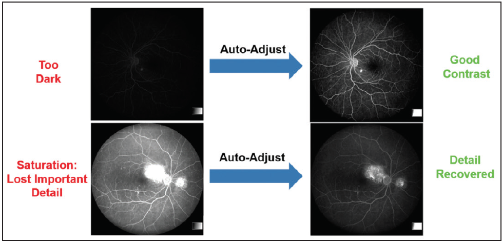
Figure 3. AutoBright technology on the CLARUS 700 adjusts images that are too dark (top row) or too saturated (bottom row) so that the imaging report is readable.
GazePoint
GazePoint technology on the CLARUS 700 leverages the power of AI to create fully detailed montage UWF images. When patient fixation is imperfect, the resulting image may be suboptimal. With GazePoint, the optic nerve becomes the point of fixation, resulting in a sharper image (Figure 4).
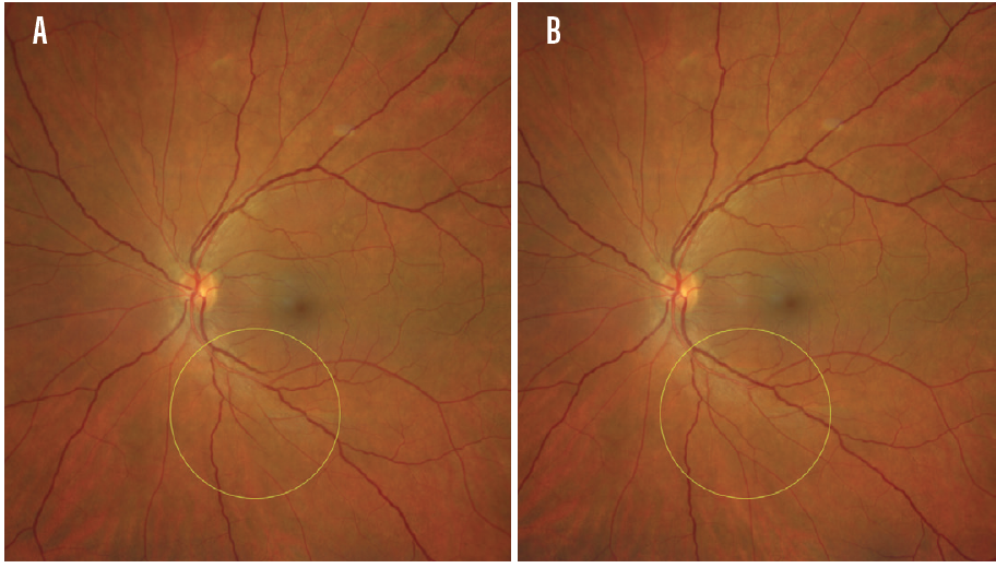
Figure 4. UWF images without perfect fixation may result in the readouts that show, for example, discontinuation of a vessel (A, yellow circle). After applying GazePoint, artificial intelligence adjusts the image, resulting in a readout that shows vessel continuation (B, yellow circle).
CASE EXAMPLE
In this patient with severe nonproliferative diabetic retinopathy, limited details are available on standard 7-field imaging (Figure 5A). When UWF imaging on FA is employed, the extent of disease and the severity of leakage are better understood (Figure 5B). With the assistance of PrecisionFocus, AutoBright, and GazePoint technologies on the CLARUS 700, this montage image is in high resolution with appropriate contrast.
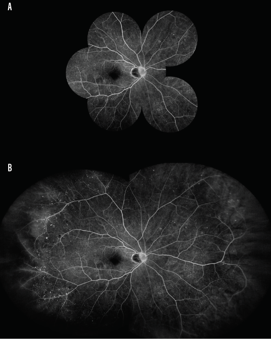
Figure 5. FA imaging of a patient with severe nonproliferative diabetic retinopathy reveals limited data when employing standard 7-field imaging (A). When UWF imaging is employed, the nature and extent of the disease is easily observed (B).
DOING MORE WITH LESS
In a busy clinic, efficiency and efficacy are key. By combining multiple modalities on a single platform in the CLARUS 700, my clinic functions productively, and my patients are able to undergo multiple tests in a short period of time. Software updates that allow sharper, more detailed images improve my clinical analysis and allow me to tailor treatments to my patients’ specific needs.
